
COSMETIC EFFICACY
Demonstrate the Efficacy of your Final Product or Ingredient
Also included: 3D pictures, macroscopic pictures, use test (self-assessment questionnaire) and dermatologically tested.
We assess the capacity of an active ingredient or end product to inhibit (whitening) or stimulate (tanning) melanin synthesis, both in clinical assays after treatment in human voluteers, and in human melanocytes and human pigmented 3D reconstituted skin (RHPE). We also analyze the functional effects on melanogenesis related genes expression regulation.
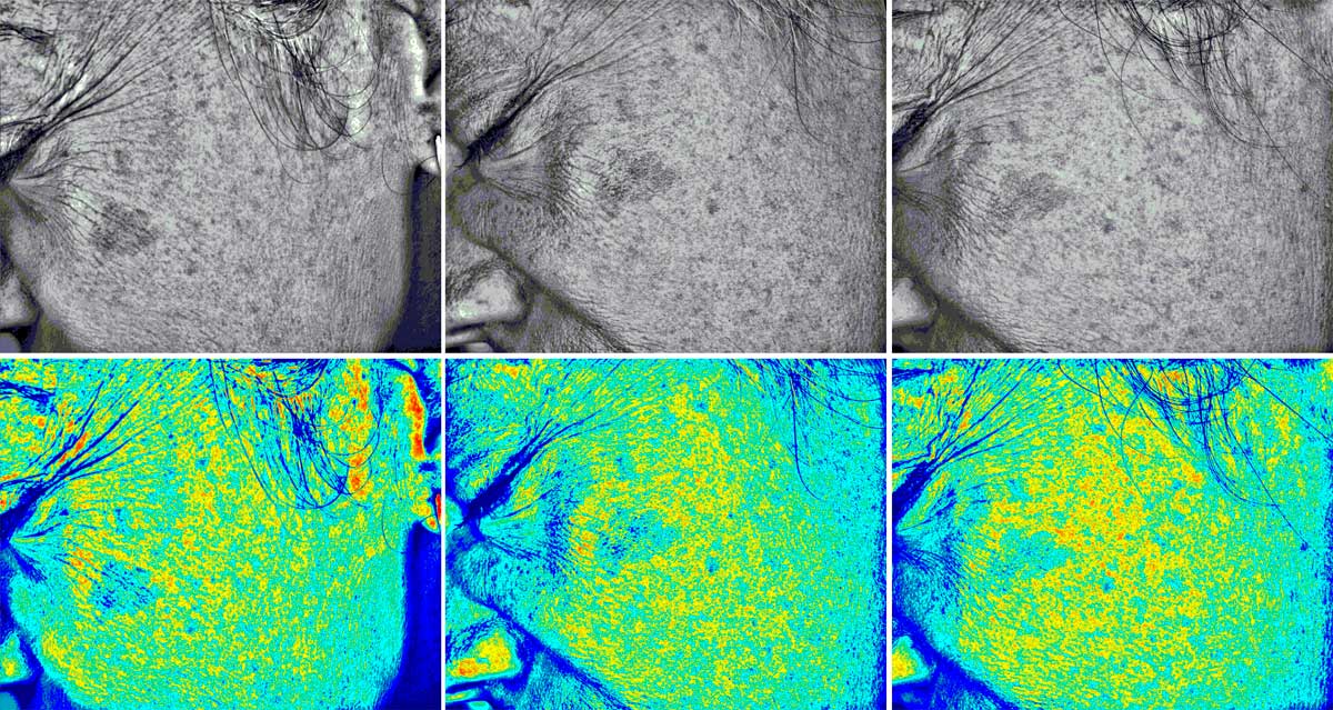
Bio Blue Light Scanner uses a high-resolution camera (5 Mpixels) and a diffuse and indirect lighting system, combined with an internal software created exclusively for the assessment of skin spots. This cutting-edge system allows us to quantify total area of dark spots, the tone of the spots, skin uniformity, skin contrast, etc.
Also included: marketing images in different formats, macroscopic pictures, use test (self-assessment questionnaire) and dermatologically tested for all volunteers.
We quantify the protective capacity of a treatment against the oxidative stress induced by urban pollution measuring reactive oxygen species (ROS) after induction with Urban Dust Particulate Matter. In the same way, we evaluate in human volunteers the dust particulates adhered to the skin after a specific exposure time.
We analyze in vitro whether an ingredient or final product protects against the oxidative stress, aging and senescence induced by ultraviolet (UV), infrared (IR) or blue light (high-energy visible, HEV) radiation, quantifying reactive oxygen species (ROS) induced after treatment of human cell lines or reconstituted 3D skin.
Similarly, we determine the protective or repairing capacity of a compound on DNA damage by quantifying thymine dimers and H2Ax activation using flow cytometry.

We assess the antiaging effects through quantification of several molecular biomarkers related to premature aging and photoaging, such as lipid peroxidation, protein carbonylation, MMPs inhibition, telomere shortening, circadian rhythms regulation and cell cycle, proteasome activation, etc.
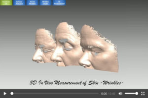
Similarly, using Bio3D Structured-light Scanner, we quantify the effects on skin sagging and neck jowls, remodeling effects on face oval, anti-rugosity in eyelids, etc. These in vivo clinical studies include 3D pictures, perfect for marketing and sales exposure, together with macroscopic pictures, use test and dermatologically tested.

We use in vitro cell cultures of Human Follicle Dermal Papilla Cells (HFDPC) to assess the capacity of a product or active ingredient to induce growth, reduce hair loss, increase hair strenght, inhibit hair graying appearance, etc.
In the same way, we use human hair follicles extracted from volunteers (ex vivo) to determine growth stimulation following Philpott’s et al., 1990 protocol. Microscopic pictures are included in this assay for each of the tested conditions.
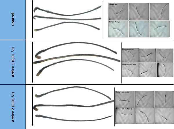
In this sense, we also analyze the efficacy of hair protectors after ex vivo topical application in human hair swatches, which we irradiate with UV radiation to generate oxidative stress and protein degradation.
Last, we use Bio Blue Light Scanner technology to determine in human volunteers the anti-hair loss and growth stimulating capacity of a treatment, together with hair graying reduction.
We determine in vitro the capacity of a compound to protect against the inflammatory response induced by an external agent such as ultraviolet radiation (UV) or bacterial lipopolysaccharide (LPS), quantifying biomarkers involved in inflammatory response and immunity like TNFa, IL-1a, IL-1b, IL-6, IL-8, IL-10, NFkB, TGFb, etc.
In the same way, we perform clinical studies on human volunteers to determine the calming and anti-redness effects of topical treatment, through subjective evaluations using questionnaires or objective quantifications using probes (redness, hydration, TEWL, etc.).

We analyze the epigenetic effects of an active ingredient or end product, through quantification of the protective effects on DNA methylation induced by an external agent, or through analysis of the effects on microRNAs expression directly involved in health and skin quality.
On the other hand, we analyze the effects on the expression of the whole human genome, through microarray analysis. We obtain genes differentially expressed and metabolic pathways mostly affected, after perform an exhaustive statistical analysis for all the tested conditions.

We assess the capacity of an active ingredient or end product to improve the skin health, both in human volunteers after topical application, as well as human dermal fibroblasts (NHDF) in vitro cultures and reconstituted 3D human skin (RHE FT).
In clinical in vivo assays with volunteers, we quantify the firmness and elasticity of the skin after a specific treatment; whereas in vitro we analyze the regulation of molecular biomarkers involved in extracellular matrix (ECM) such as collagen, elastin, matrix metalloproteinases, fibronectin, laminin, versican, lumican, etc., both at gene expression level using RT-qPCR and protein level using ELISA.
At Bionos, we determine in vitro the wound-healing and regenerating capacity of a product through the Wound-Healing or Scratch Assay, after treatment in human cell lines. The application of a new image technology allow us to obtain at the same time a time-lapse movie, perfect to the demonstration of the effect in marketing and sales.
On the other hand, we assess the cytotoxic and proliferative capacity of a determine active ingredient, after cell viability evaluation through MTT assay, in different conditions of culture.
At the same time, we assess the regenerative capacity in human volunteers after topical application in forearm and quantification using specific probes (mexameter, etc.).

Using confocal microscopy, we determine the penetration capacity of a specific compound in cutaneous surface after application in 3D human reconstituted skin (RHE), pig skin or human cell lines.
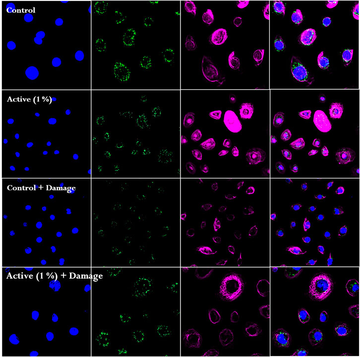
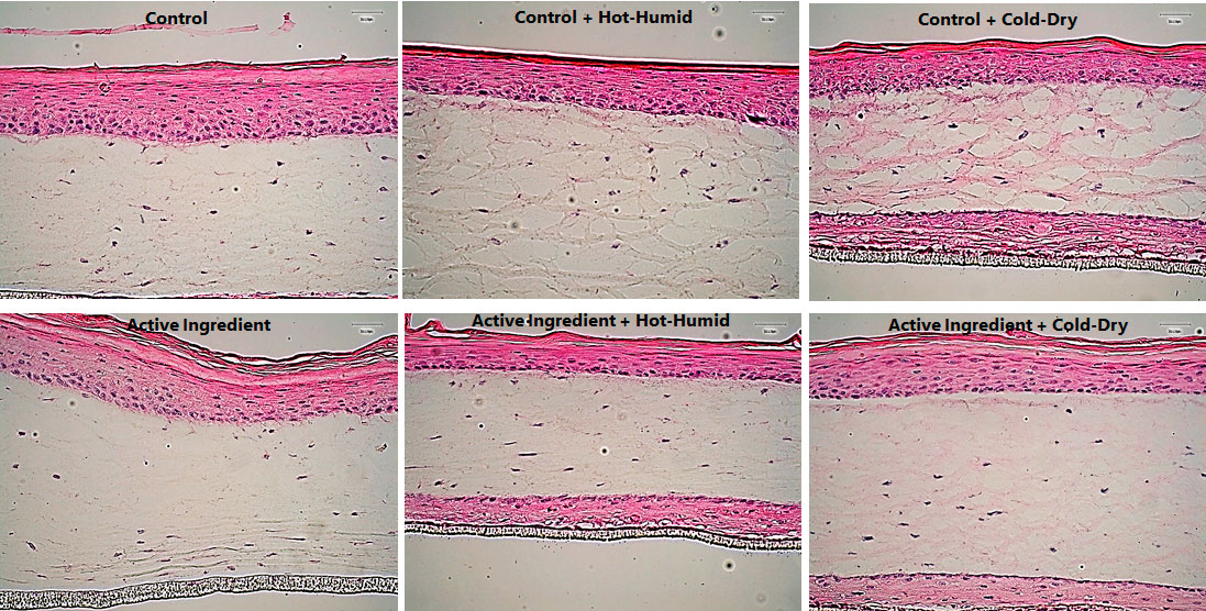
In the same way, we assess the functionality of different compounds through immunostaining techniques, using different stainings (DAPI, fluorescence, hematoxylin-eosin, phalloidin, PCNA, etc.) and specific antibodies (NRF2, ROS, apoptosis, etc.).
TEST YOUR PRODUCTS
Do you have any questions or doubts?
Fill in the contact form and we will reply as soon as possible.
CALL US
+34 96 124 32 19
CONTACT
bionos@bionos.es
VISIT US
Av. Fernando Abril Martorell, 106.
Hospital La Fe, Torre A, Planta 1.
46026 Valencia